Cardiovascular Posters
Poster: Cardiovascular Posters
18 - A case of Persistent Left Superior Vena Cava with an attenuated left-sided azygos vein accompanying normal right-sided antimeres
Sunday, March 24, 2024
5:00pm - 7:00pm US EDT
Location: Sheraton Hall
Poster Board Number: 18
There are separate poster presentation times for odd and even posters.
Odd poster #s – first hour
Even poster #s – second hour
Co-authors:
There are separate poster presentation times for odd and even posters.
Odd poster #s – first hour
Even poster #s – second hour
Co-authors:
Jeremy Rosenbaum - Medical Student, Keck School of Medicine of the University of Southern California; Robert Raad - Medical Student, Keck School of Medicine of the University of Southern California; Michelle Dong - Medical Student, Keck School of Medicine of the University of Southern California; Brandon Hsiao - Medical Student, Keck School of Medicine of the University of Southern California; Hannah Nash - Medical Student, Keck School of Medicine of the University of Southern California; Isabella Smith - Medical Student, Keck School of Medicine of the University of Southern California; Ajay Prasad - Medical Student, Division of Acute Care Surgery, Keck School of Medicine of the University of Southern California; Anaar Siletz, M.D., Ph.D. - Assistant Professor of Clinical Surgery, Division of Acute Care Surgery, Keck School of Medicine of the University of Southern California; Biren Patel, Ph.D. - Professor, Integrative Anatomical Sciences, Keck School of Medicine of the University of Southern California; Tea Jashashvili, M.D., Ph.D. - Assistant Professor, Integrative Anatomical Sciences, Keck School of Medicine of the University of Southern California; Kristian Carlson, Ph.D. - Professor, Integrative Anatomical Sciences, Keck School of Medicine of the University of Southern California

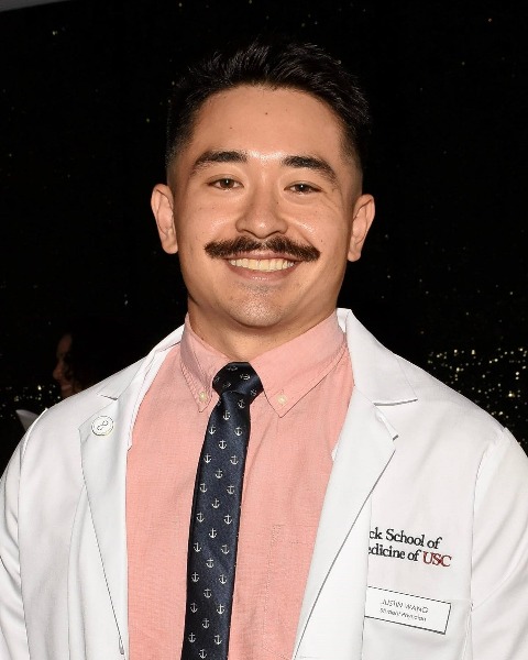
Justin L. Wang, MS
Medical Student
Keck School of Medicine of USC
Los Angeles, California, United States
Presenting Author(s)
Abstract Body : A persistent left superior vena cava (PLSVC) occurs as a developmental phenomenon, often when the left cardinal vein does not fully degenerate. A PLSVC is reportedly the most common thoracic venous anomaly; it is typically asymptomatic and undiagnosed and is estimated to be prevalent in 0.2 – 3% of the overall population. In adults, this is often represented as an extrapericardial vein, via the left brachiocephalic, that drains into the right atrium by either joining the coronary sinus or paralleling its path creating a duplicate drainage. A PLSVC has been reported to co-occur with a right superior vena cava (RSVC) or persist alone, and it may be accompanied by a left-sided azygos vein and a right-sided hemiazygos vein. Here we document bilateral superior vena cavae (BSVC) and azygos veins. The heart and mediastinum were dissected from a 95-year-old man who died of diastolic congestive heart failure and donated his remains to a medical gross anatomy course. Vessel diameters were measured with calipers. Photographs were taken, and a 3D model was created using photogrammetry.
Although the left-sided azygos vein was reduced in size, it appeared functional and mirrored the arrangement of the larger right-sided azygos vein. Neither the RSVC nor the right-sided azygos vein exhibited abnormalities. Comparative blood vessel diameters between the right and left azygos veins suggest that blood from the lower left quadrant of the thorax primarily drained into the right-sided azygos vein by crossing the posterior mediastinum via a well-developed hemiazygos vein. The individual also exhibited a seldom-reported variation in which the coronary sinus and posterior interventricular vein drained directly into the PLSVC via a single lateral entrance into the right atrium. The PLSVC was dilated to a diameter of 16.4 mm at its greatest width.
A PLSVC is often discovered incidentally in clinical practice after radiological imaging. Numerous variations have been identified, and their direct health implications can range from asymptomatic to life-threatening, although the latter is less common to observe in adults. Prior awareness of vascular anatomical variants is particularly important in trauma and emergency surgery, when preoperative imaging may be impossible. The present case of anatomical variation is instructive since the reduced nature of the PLSVC and its accompanying left azygos vein draining into the right atrium may be more challenging to diagnose through radiological imaging. Physicians should be cognizant of this spectrum of size variation, rather than a presence-absence condition, prior to vascular intervention.

