Upper Limb Case & Anatomical Studies Posters
Poster: Upper Limb Case & Anatomical Studies Posters
61 - Subscapular Artery Variations and Military Personnel Considerations - A Case Report and Review
Sunday, March 24, 2024
5:00pm - 7:00pm US EDT
Location: Sheraton Hall
Poster Board Number: 61
There are separate poster presentation times for odd and even posters.
Odd poster #s – first hour
Even poster #s – second hour
Co-authors:
There are separate poster presentation times for odd and even posters.
Odd poster #s – first hour
Even poster #s – second hour
Co-authors:
Amy Pearson - Medical Student, School of Medicine, Uniformed Services University of Health Sciences; Guinevere Granite, MS, MA, PhD - Director of Human Anatomy, Department of Surgery, Uniformed Services University of Health Sciences; Maria Leighton, M.D. - Assistant Professor, Department of Surgery, Uniformed Services University of Health Sciences; Jinbum Dupont, M.S. - Medical Student, School of Medicine, Uniformed Services University of Health Sciences; Elizabeth Maynes, M.D. - Assistant Professor, Department of Surgery, Uniformed Services University of Health Sciences
.jpg)
Olivia J. Staser
Medical Student
Uniformed Services University of Health Sciences
Bethesda, Maryland, United States
Presenting Author(s)
Abstract Body : Introduction: The axillary artery provides blood to the upper limb and thoracic region. It contains three subdivisions with six branching vessels, starting where the subclavian artery ends, distal to the lateral border of the first rib. The classic presentation is uncommonly found in approximately 10% of patients. Investigation into variation patterns can help elucidate regional anatomy for surgical and medical management.
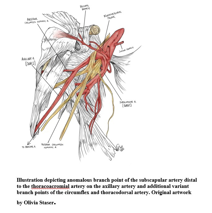
Methods and Case Description: During a routine dissection of a 97-year-old white female cadaver, an unusual branching pattern of the axillary artery was identified. The subscapular artery branched off just distal to the junctional point of the thoracoacromial artery in axillary artery Section 2. The subscapular artery traveled superficially to the medial cord and ulnar nerve and contained the branch point of the posterior circumflex artery. The subscapular artery then gave rise to the circumflex scapular artery and thoracodorsal artery. The axillary artery then dove underneath the lateral cord of the brachial plexus and passed over the musculocutaneous nerve and continued along the standard pathway.

Significance and Implication: Axillary artery variations add technical complication and increased risk during surgery, such as treating aortic dissection, aortic aneurysm, or performing coronary artery bypass. Injury to the axillary artery can occur at a rate of 3-9% in military personnel. Knowledge of axillary artery variations is significant for radiological imaging assessments and accurate interpretation of the region. In the case presented, we hypothesize that the individual would have been at risk for medial cord involvement in the event of vascular expansion due to the novel pinch-point introduced by the anomalous subscapular branch point.
Conclusion: The location and juxtaposition of neurovascular structures of the axillary region create challenges for surgeons faced with treating injuries to the area. Axillary artery variations have implications in surgical and radiological contexts and are particularly important in the military. Therefore, knowledge of pertinent variations of the region is vital since time will be a limiting factor affecting upper limb structure morbidity and overall surgical outcomes.
Methods and Case Description: During a routine dissection of a 97-year-old white female cadaver, an unusual branching pattern of the axillary artery was identified. The subscapular artery branched off just distal to the junctional point of the thoracoacromial artery in axillary artery Section 2. The subscapular artery traveled superficially to the medial cord and ulnar nerve and contained the branch point of the posterior circumflex artery. The subscapular artery then gave rise to the circumflex scapular artery and thoracodorsal artery. The axillary artery then dove underneath the lateral cord of the brachial plexus and passed over the musculocutaneous nerve and continued along the standard pathway.
Significance and Implication: Axillary artery variations add technical complication and increased risk during surgery, such as treating aortic dissection, aortic aneurysm, or performing coronary artery bypass. Injury to the axillary artery can occur at a rate of 3-9% in military personnel. Knowledge of axillary artery variations is significant for radiological imaging assessments and accurate interpretation of the region. In the case presented, we hypothesize that the individual would have been at risk for medial cord involvement in the event of vascular expansion due to the novel pinch-point introduced by the anomalous subscapular branch point.
Conclusion: The location and juxtaposition of neurovascular structures of the axillary region create challenges for surgeons faced with treating injuries to the area. Axillary artery variations have implications in surgical and radiological contexts and are particularly important in the military. Therefore, knowledge of pertinent variations of the region is vital since time will be a limiting factor affecting upper limb structure morbidity and overall surgical outcomes.

